Decoding Osteoarthritis: Choosing Between X-Rays and MRI for Accurate Diagnosis
X-Rays: The Traditional Approach
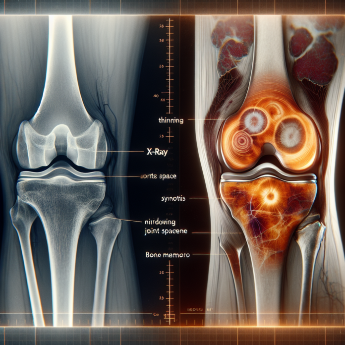
X-rays have long been used as a go-to diagnostic tool for osteoarthritis. They provide a clear image of the bone structures and are capable of showing changes that occur within the joints. X-rays are particularly adept at revealing:
- Bone Spurs: Bone spurs, or osteophytes, can be easily visible on X-rays. These bony projections tend to form along joint margins and are a hallmark sign of osteoarthritis.
- Joint Space Narrowing: As cartilage wears away, the joint space between bones narrows. X-rays performed under load-bearing conditions, such as standing weight-bearing images, can show this narrowing, indicating the extent of cartilage loss and the severity of osteoarthritis.
- Bone-on-Bone Contact: In advanced stages of osteoarthritis, the cartilage may be completely worn away, leading to bone-on-bone contact. This is often clearly depicted on X-rays taken while the patient is standing, as the compression from body weight can highlight the joint integrity (or lack thereof).
MRI: The Advanced Imaging Choice
While X-rays focus on bones, MRI scans provide a comprehensive look at all joint components. MRI is especially valuable for viewing:
- Articular Cartilage: MRI can showcase the quality and thickness of the cartilage, highlighting areas where it may be worn thin or where lesions and holes exist. This can provide a more nuanced understanding of the cartilage’s condition compared to an X-ray.
- Soft Tissue Structures: Unlike X-rays, MRIs can visualize soft tissues such as ligaments, tendons, and the menisci. This is vital for diagnosing complex joint issues that may accompany or exacerbate osteoarthritis.
- Subchondral Bone Changes: MRI can detect changes in the subchondral bone, such as edema or cyst formation, which are not visible on X-rays and could be key indicators of early osteoarthritis or related joint pathologies.
Comparative Advantages and Limitations
While both tools offer valuable insights, each has its limitations. X-rays, due to their focus on bones, cannot provide information about the soft tissues or the cartilage itself. However, the weight-bearing X-ray can give a more accurate representation of the joint’s functional state because the patient is in a natural standing position, showing the true effects of weight and gravity on the joint.
Conversely, MRI does not have the capacity to show real-time weight-bearing conditions, as the examination is typically performed while lying down. This could potentially under-represent the severity of the condition due to absence of load-bearing forces during imaging.
Beyond Imaging: Comprehensive Treatment Decisions
The decision to opt for a joint replacement is far more complex and patient-specific than simply interpreting images. While imaging provides a picture of the joint condition, it must be considered alongside clinical assessments such as:
- Pain Levels: The degree of discomfort and how it affects daily living activities is a key consideration. Some patients with significant image-based changes may experience minimal symptoms, while others with moderate changes might endure substantial pain.
- Functional Impact: The impairment in daily activities, mobility, and quality of life are crucial factors. If osteoarthritis limits a person’s functionality significantly, more aggressive interventions might be considered.
- Previous Treatments: Prior attempts to manage the condition, such as physiotherapy, medication, or injections, need to be considered when deciding the next step in treatment.
Therefore, while imaging is critical, the decision for procedures like joint replacement should be based on a combination of imaging results, clinical assessment, and patient-specific considerations.
In conclusion, both X-rays and MRI scans are indispensable tools in the diagnosis and management of osteoarthritis. The choice between the two largely depends on the specific needs of the patient, the details of the condition, and how significantly the condition affects the patient’s life. When used judiciously in combination with a thorough clinical evaluation, these imaging modalities can guide effective treatment planning, tailored to each patient’s unique circumstances.
X-rays and MRI scans both aid in diagnosing osteoarthritis, each with unique strengths. X-rays show bone structure changes, while MRI offers detailed views of cartilage and soft tissues.
