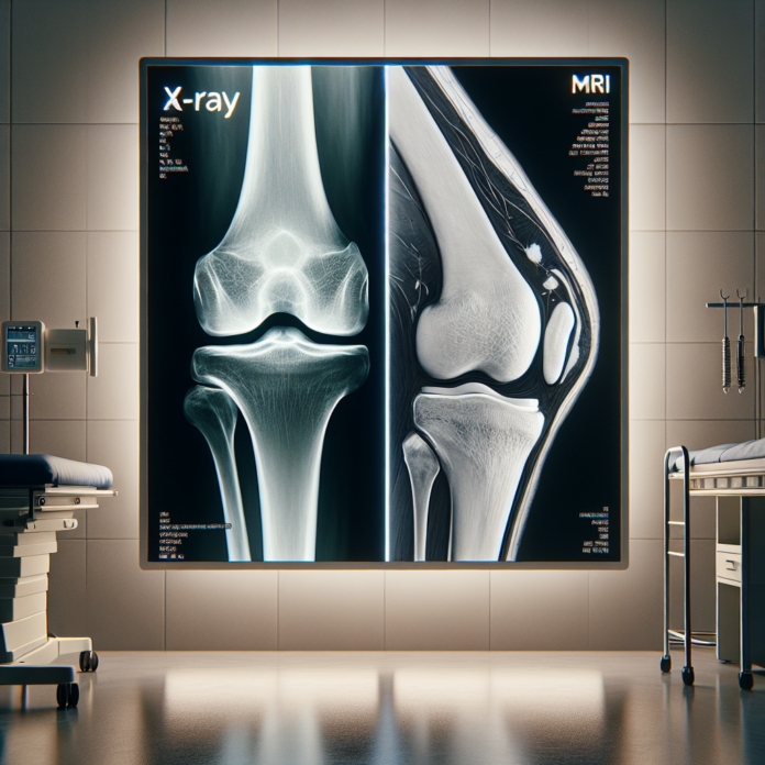
Osteoarthritis is a common joint condition that can significantly impact one’s quality of life, and accurately diagnosing its presence and severity is crucial for effective management. In today’s blog, we delve into the ongoing debate between the use of X-rays and MRIs to detect and assess osteoarthritis. Drawing insights from Dr. David Guyer, a triple board-certified orthopedic surgeon, we’ll explore the pros and cons of each imaging technique, helping you make informed decisions about your joint health. Whether you’re a patient wondering about your treatment options or simply curious about the latest advancements in medical imaging, this blog will provide you with a comprehensive understanding of how X-rays and MRIs play a role in the diagnosis and evaluation of osteoarthritis. Stay tuned to learn about the unique capabilities of each method, and how they can be utilized to tailor a personalized approach to managing your joint health.
X-rays: Simple Yet Effective
X-rays are a staple in the world of medical imaging due to their ability to paint a clear picture of the bone structure. They excel in visualizing bones and can effectively show the bone spacing that indicates the presence of cartilage. This characteristic makes X-rays particularly useful in identifying osteoarthritis, as it often manifests as a narrowing of the joint space due to cartilage loss. Here’s what X-rays can show when assessing osteoarthritis:
- Bone Structure: X-rays provide a detailed view of the bones, making it easier to identify deformities and the development of bone spurs—an indicator of advanced osteoarthritis.
- Joint Space Narrowing: The space between bones on an X-ray is a proxy for cartilage thickness. A narrowing of this space signals cartilage wear, a hallmark of osteoarthritis.
- Bone on Bone Contact: Weight-bearing X-rays, performed in many orthopedic offices, can show the extent of bone on bone contact. This is a crucial indicator of the severity of arthritis, which can be better visualized under the influence of gravity and body weight.
However, X-rays have limitations. They only show the bones and do not provide information about the meniscus, ligaments, or detailed cartilage condition.
MRIs: A Comprehensive View
Magnetic Resonance Imaging (MRI), on the other hand, offers a more comprehensive view of the joint’s internal structures. This includes not just the bones, but also the soft tissues like cartilage, ligaments, and tendons. An MRI can detail the specific areas where cartilage may be thinning or has been breached, offering a comprehensive assessment of joint health. Here’s what MRIs add to the diagnosis of osteoarthritis:
- Cartilage Condition: MRIs can visualize the thickness and health of articular cartilage, revealing specific areas of thinning or damage that X-rays cannot show.
- Soft Tissue Evaluation: They provide detailed images of all soft tissue components within the joint, such as ligaments and tendons, helping to identify any concurrent injuries or conditions.
- Detailed Assessment Without Weight Influence: While lying down during an MRI might underestimate arthritis severity due to the absence of body weight, it can nonetheless reveal the presence of minute cartilage defects and other abnormalities.
Choosing Between X-rays and MRI
The choice between X-rays and MRIs should be informed by the specific details of each patient’s condition. While MRIs provide a detailed picture that sketches the condition of the cartilage and other soft tissues, X-rays highlight the alignment, structural stability, and bone changes more prominently, especially under weight-bearing conditions.
- Consider X-rays if: You need a quick, cost-effective method for assessing bone health and joint space. It’s also useful if you seek to understand how gravity affects joint spacing or if cost and availability are concerns.
- Consider MRI if: You need a detailed evaluation of the soft tissues, have complex symptoms that are not fully explained by bone conditions, or when there is a need for a comprehensive overview of the joint’s internal condition.
Beyond Imaging: Comprehensive Care
Ultimately, while imaging studies are indispensable in diagnosing osteoarthritis, they are just one piece of the puzzle. A crucial aspect of treatment decisions involves the patient’s level of pain, physical activity limitations, and response to previous treatments. A clinician considers all these factors before determining whether surgical intervention, such as a joint replacement, is necessary.
Both imaging methods have their strengths and roles in managing osteoarthritis. By understanding these roles, patients can have a more meaningful dialogue with their healthcare providers and tailor a personalized care plan that fully addresses their specific needs and lifestyle goals.
Stay informed about your joint health, and consult professional healthcare advice to make the best decisions for your unique situation. Whether dealing with the onset of symptoms or managing long-standing osteoarthritis, understanding the imaging tools at your disposal is a powerful step in achieving effective treatment outcomes.
