“`html
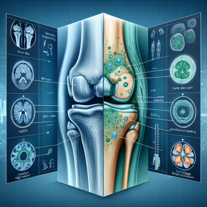
When considering the diagnosis and management of osteoarthritis, both X-rays and MRIs offer valuable insights, though each provides a unique perspective. Understanding the capabilities and limitations of these imaging techniques can aid in making informed decisions regarding treatment paths.
X-Rays: A Traditional Approach
X-rays are often the first step in diagnosing osteoarthritis. They are particularly useful for visualizing bones and the joint space, offering a clear assessment of bone health. Here are some key advantages:
- Bone Visualization: X-rays effectively show changes in bone structure. This includes the presence of bone spurs, which are bony projections that develop along the edges of bones and are common in osteoarthritis.
- Joint Space Evaluation: The space between bones, visible on an X-ray, can indicate cartilage loss. A reduced joint space typically suggests cartilage wear, a hallmark of osteoarthritis.
- Weightbearing Insight: When X-rays are taken while a patient is standing, or weightbearing, they provide an indication of how the joint functions under pressure. This can highlight the severity of arthritis by showing direct bone-on-bone contact when weight is applied.
However, X-rays have their limitations. They do not reveal the condition of soft tissues such as cartilage, ligaments, or tendons. This can be a significant drawback, as osteoarthritis affects not just the bone but the joint as a whole.
MRI: A Comprehensive View
Magnetic Resonance Imaging (MRI) offers a more detailed view of the joint, encompassing both hard and soft tissue structures:
- Soft Tissue Assessment: MRIs can visualize the articular cartilage, allowing for an evaluation of its thickness and integrity. This is crucial in determining the extent of cartilage damage.
- Detailed Imaging: MRI can show subtle changes such as cartilage thinning, small tears, or other degenerative signs that X-rays cannot detect. It can display the condition of ligaments and tendons, offering a comprehensive view of the joint’s health.
- Three-Dimensional Insight: Unlike the two-dimensional image from an X-ray, an MRI provides a three-dimensional perspective, enhancing the understanding of the joint’s condition.
Nonetheless, MRIs are not without their challenges. The primary concern with MRI is that the imaging is typically done with the patient lying down. This removes the effect of gravity and body weight on the joint, which can sometimes underestimate the severity of arthritis. Additionally, MRIs are more expensive and less readily available than X-rays.
Choosing Between X-Rays and MRI
The decision between X-rays and MRI is not always straightforward. Both have their roles depending on the clinical situation:
- Initial Assessment: X-rays are generally favored for initial assessments due to their ability to quickly identify significant bone changes and joint space narrowing, which are indicative of advanced osteoarthritis.
- Complex Cases: When further detail is needed, or when symptoms do not correlate with X-ray findings, an MRI might be warranted. This could be the case if there is suspicion of soft tissue involvement, or if there is a need to explore cartilage condition in detail.
It’s important to highlight that neither imaging modality alone can dictate treatment decisions, such as the need for joint replacement. These decisions are multifaceted, relying on a combination of imaging results, the severity of symptoms, the impact on quality of life, and the success or failure of previous treatments.
Additional Considerations
- Patient Experience: MRI machines can be claustrophobic for some, which may be a consideration for patients who are apprehensive about enclosed spaces.
- Availability and Cost: X-rays are more accessible and cost-effective, making them a practical first-line choice in many cases.
Ultimately, the choice between X-rays and MRI should be guided by a comprehensive evaluation by a healthcare professional. Each case is unique, and factors such as patient age, activity level, and overall health should be considered in the decision-making process.
For those seeking more comprehensive guidance, Dr. David Guyer offers an eBook titled “The Arthritis Solution,” which provides detailed insights into managing osteoarthritis. Additionally, exploring options beyond surgery, such as regenerative medicine, may offer alternative pathways to relief and improved function.
Understanding the role of X-rays and MRI in diagnosing and monitoring osteoarthritis can empower patients to engage in their healthcare journey proactively. In partnership with skilled orthopedic specialists, patients can strive towards optimal joint health and enhanced quality of life.
“`
