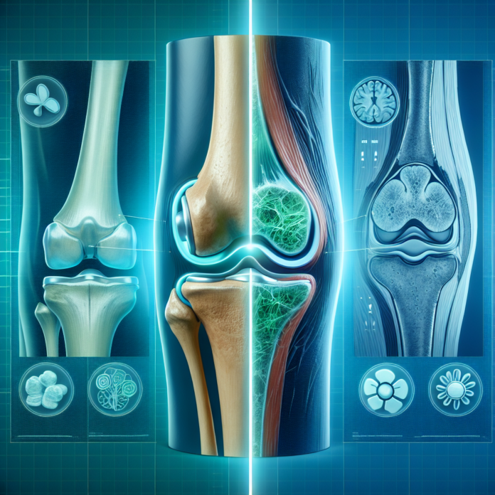
When it comes to understanding the complexities of osteoarthritis, both patients and practitioners often find themselves navigating the nuances of various diagnostic tools. In the realm of medical imaging, X-rays and MRIs stand out as two pivotal options, each offering distinct insights into the condition and its severity. Dr. David Guyer, a renowned orthopedic surgeon, delves into this very topic in a recent video, exploring the pros and cons of these imaging techniques. His expertise in sports medicine, anti-aging, and regenerative medicine brings a comprehensive perspective to the discussion, helping viewers decipher whether an X-ray or an MRI is more suited for their needs. As we expand upon his insights here, we’ll examine the capabilities and limitations of these diagnostic tools, ultimately guiding you through making an informed decision for your osteoarthritis management.
X-rays: The Basics
X-rays are often the first line of imaging used when osteoarthritis is suspected. They provide a clear picture of the bones and the spacing between them. This is significant because osteoarthritis is characterized by the degeneration of cartilage, the cushiony material that helps our joints move smoothly. When cartilage wears away, the space between bones decreases, and X-rays can effectively show this “bone-on-bone” state.
Pros of X-rays:
- Visualization of Bone Structure: X-rays are excellent for viewing the structural alignment of bones. This is particularly useful in identifying bone spurs – bony projections that form along joint margins and are common in osteoarthritis.
- Cost-effectiveness: X-rays are generally less expensive than MRIs, making them a more affordable option for an initial assessment.
- Accessibility: Most orthopedic offices are equipped to perform X-rays, which means they can be done quickly and easily during a routine visit.
Cons of X-rays:
- Limited Soft Tissue Details: X-rays do not show the soft tissues such as cartilage, ligaments, and tendons. Therefore, they cannot provide detailed information about the condition of these structures.
- Less Sensitivity in Early Detection: X-rays may not detect the early stages of osteoarthritis as effectively as MRIs, since significant changes in bone structure may take time to develop.
MRIs: A Closer Look
MRI, or Magnetic Resonance Imaging, offers a more comprehensive look at the joint structures, providing detailed images of both bones and soft tissues. An MRI can reveal more about the condition of the cartilage, ligaments, and tendons, giving a fuller picture of the overall joint health.
Pros of MRIs:
- Detailed Imaging of Soft Tissues: MRIs are superior in assessing the condition of cartilage, allowing the visualization of thinning or tears, and damage to surrounding ligaments and tendons.
- Early Detection: An MRI can detect early degenerative changes in the joint, often before changes can be seen on X-rays.
Cons of MRIs:
- Cost and Accessibility: MRIs are more expensive and time-consuming compared to X-rays. They may also not be as readily accessible due to the need for specialized equipment.
- Misleading Results Underweight: Because MRI scans are performed while the patient is lying down, they may not accurately demonstrate how the joint behaves under normal weight-bearing conditions, potentially leading to a misestimation of the severity of the arthritis.
Determining the Need for Joint Replacement
While imaging tests play a foundational role in diagnosing osteoarthritis, they are not the sole determinants for deciding on a joint replacement surgery. The decision is multifactorial, involving:
- Severity of Symptoms: A patient’s pain level, mobility issues, and how these factors impact daily life hold immense weight in treatment decisions.
- Previous Treatments: Considering past interventions and their efficacy is crucial. These can include physical therapy, injections, or medications.
- Patient Activity Level and Goals: A patient’s lifestyle, activity level, and personal goals are also important factors in deciding whether to pursue more invasive treatment options like joint replacement.
Both X-rays and MRIs provide valuable information about osteoarthritis, but neither can independently dictate the need for surgery. These imaging tests can offer guidance, but they should be considered alongside clinical evaluation and patient history to reach a decision that best aligns with the patient’s needs and preferences.
In conclusion, choosing between an X-ray or MRI depends largely on the specifics of the case at hand. An X-ray may suffice for a general assessment, especially in more advanced stages of osteoarthritis, while an MRI can provide a deeper understanding of complex or early-stage conditions. Consulting with a specialized healthcare provider can help navigate these options, ensuring that the chosen path aligns with both the clinical findings and the holistic needs of the patient. Whether you are dealing with osteoarthritis or any other orthopedic issues, understanding the nuances of these diagnostic tools is instrumental in crafting a comprehensive and effective care plan.
