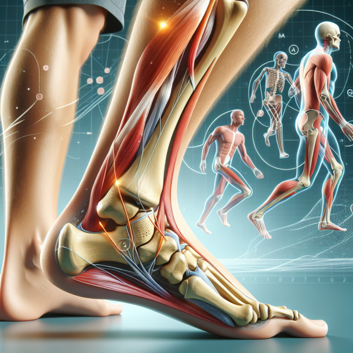
Peroneal tendon subluxations are a complex condition typically following lateral ankle sprains. The peroneal tendons, located behind the fibula on the outer side of the ankle, play a crucial role in stabilizing the foot and aiding in ankle movement. Unfortunately, these tendons can become a source of significant discomfort and dysfunction when the tissue holding them, the retinaculum, is torn or stretched. This tearing usually occurs during an inversion injury, where the ankle twists inward, placing immense stress on the structures on the outer side of the ankle, including the retinaculum.
The Diagnostic Challenge
The phenomenon of peroneal tendon subluxation presents a unique challenge both in terms of diagnosis and treatment. At the onset, the clicking or slipping sensation in the foot during movement, such as eversion (turning the foot outward), is often the first indication for patients. This can be confusing when MRI scans, considered the gold standard for soft tissue imaging, return inconclusive results. The failure of an MRI to capture a clear picture of the damage often arises from improper imaging planes or the subtle nature of the tear, which eludes even the most skilled radiologists.
This diagnostic conundrum raises the question of alternative imaging methods. Dynamic ultrasound emerges as a viable option in this scenario. Unlike static MRIs, dynamic ultrasound allows real-time observation of the tendons as the foot moves, providing a clearer picture of any subluxation occurring and thus confirming the diagnosis. However, a thorough physical examination remains pivotal. An orthopedic specialist can often diagnose peroneal tendon subluxation based on symptoms and physical signs alone, bypassing the need for imaging altogether in certain cases.
When Surgery Becomes Necessary
When addressing treatment, it’s imperative to understand that surgery becomes a necessity in most scenarios of peroneal tendon subluxation, especially if the tendons are fully subluxing. A torn retinaculum means that these tendons have lost their stabilizing support, leading to friction against the fibula, which can cause further fraying and eventual rupture. This progressive damage can exacerbate pain and diminish ankle function, making surgical intervention imperative to restore stability and prevent long-term complications.
Non-surgical treatments, including physical therapy, taping, or regenerative medicine techniques like platelet-rich plasma (PRP) injections, unfortunately, offer limited efficacy in cases where the retinaculum is torn. These methods may provide temporary relief but typically fail to address the underlying structural instability, which is essential for lasting recovery.
The Surgical Approach
Surgical repair aims to restore the integrity of the retinaculum and reposition the tendons into their natural groove, allowing them to function correctly without slipping. The surgery’s specifics depend on the severity of the tendon displacement and any accompanying ankle damage, which can often be identified via a pre-operative MRI. This imaging helps in planning the surgical approach, ensuring any additional injuries are addressed concurrently.
Consultation and Decision-Making
It’s understandable that the prospect of surgery may be daunting, particularly for patients with an aversion to invasive procedures. However, delaying or avoiding necessary surgery can lead to prolonged discomfort, further tendon damage, and ultimately more complex surgical procedures down the line. Therefore, consulting with a specialized orthopedic surgeon is crucial to weigh the risks and benefits and to proceed with the most appropriate treatment plan based on individual symptoms and lifestyle needs.
Conclusion
In summary, peroneal tendon subluxation following an ankle sprain is a condition that requires careful diagnostic and therapeutic considerations. Accurate diagnosis often necessitates a combination of dynamic imaging and expert clinical assessment, while effective treatment frequently hinges on timely surgical intervention. Although the journey from injury to recovery can be challenging, understanding the nature of the injury and the available treatment options can empower patients to make informed decisions and recover more effectively.
