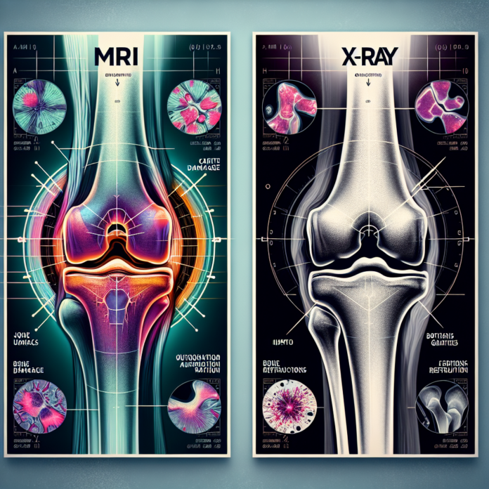
When it comes to diagnosing osteoarthritis, the debate between using X-rays or MRI scans is a common one. As a triple board-certified orthopedic surgeon and a specialist in space medicine, anti-aging, and regenerative medicine, Dr. David Guyer frequently addresses this question in his educational video series. While X-rays offer a clear view of the bones and the spaces between them, highlighting any bone spurs or signs of bone-on-bone contact, MRIs provide a detailed picture of the cartilage, ligaments, and tendons, allowing for a comprehensive assessment of joint health. Both diagnostic tools have their merits and drawbacks, and understanding these can help you make informed decisions about the management of osteoarthritis. In this blog, we delve deeper into the advantages and limitations of each method and explore how they contribute to a holistic understanding of arthritis severity, shedding light on why neither should solely dictate the need for joint replacement surgery.
X-Rays: The Traditional Approach
X-rays have long been the cornerstone of diagnosing osteoarthritis. They are particularly adept at displaying:
- Bone Structures: X-rays clearly show the bone anatomy, including any changes such as bone spurs, which are growths that can develop as arthritis progresses.
- Joint Space: The images highlight the space between bones, an indirect indicator of cartilage health. As osteoarthritis advances, this space typically narrows, suggesting cartilage loss.
- Bone-On-Bone Contact: When the cushioning cartilage wears down completely, bones may rub directly against each other, a condition easily seen in an X-ray.
Despite their clear imagery of bone-related changes, X-rays do have their limitations. They cannot visualize soft tissues like cartilage, ligaments, or tendons, making it challenging to assess early-stage arthritis where cartilage damage has begun but bone changes are not yet significant.
MRI: A Comprehensive Look
Magnetic Resonance Imaging (MRI) offers a more complete view of the joint:
- Cartilage Visualization: MRI can directly visualize cartilage, detecting thinning, tears, or even tiny defects that might not yet influence the bone.
- Soft Tissue Insight: This imaging method also provides detailed images of ligaments, tendons, and muscles, giving a comprehensive overview of the joint’s condition.
- Early Detection: Because MRIs can identify soft tissue changes, they can be instrumental in diagnosing osteoarthritis before bone changes occur.
However, MRIs also come with their drawbacks. One particular challenge is that the images are taken without the influence of gravity or body weight, which can sometimes lead to an underestimation of arthritis severity. For instance, lying down during an MRI might mask how compression affects the joint when weight-bearing.
Weight-Bearing Considerations
Weight-bearing X-rays can add value by simulating the effects of gravity on the joint. When standing, the joint is under natural stress, which can highlight bone-on-bone contact more clearly than when the body is at rest. This approach provides insight into how arthritis impacts function and can offer a clearer picture of joint degradation under typical conditions.
Interpreting Results for Treatment
Neither X-rays nor MRIs alone dictate the necessity of a surgical intervention like a knee or hip replacement. These imaging tools are part of the broader diagnostic picture, which includes:
- Symptom Severity: The level of pain and how it affects daily life are crucial factors. A person with severe X-ray findings might manage well with minimal symptoms, while someone with moderate imaging results could experience debilitating pain.
- Treatment History: Previous treatments and their effectiveness play into decision-making. For instance, if conservative therapies like physical therapy, medication, or injections have failed, surgery might be more strongly considered.
- Functional Impact: The extent to which osteoarthritis limits mobility and daily activities often weighs heavily in treatment decisions.
A Holistic Approach to Decision Making
Ultimately, the choice between using X-rays or MRIs—or a combination of both—depends on the individual case. An orthopedic surgeon might opt for X-rays initially to gauge general bone health and then utilize an MRI to delve deeper into soft tissue concerns.
The key takeaway is that while imaging plays a crucial role in diagnosing osteoarthritis, it is not the sole factor in treatment decisions. A collaborative approach, involving patients and healthcare providers in discussing symptoms, lifestyle impacts, and imaging results, leads to the most personalized and effective treatment strategy.
For patients seeking more comprehensive information, resources like Dr. Guyer’s e-book, “The Arthritis Solution,” can provide valuable insights into managing osteoarthritis beyond imaging. In a world where technology continues to evolve, keeping informed and considering multiple perspectives ensures the best possible outcomes in the fight against osteoarthritis.
