In the realm of orthopedic injuries, a fracture of the medial femoral condyle stands out as a particularly uncommon affliction, especially among adults. This rare occurrence is typically seen in children and often results from a significant traumatic force. In this blog, we delve into the intricacies of this type of fracture, when surgical intervention becomes necessary, and what the journey to recovery looks like. Drawing insights from Dr. David Guyer, a triple board-certified orthopedic surgeon, sports medicine specialist, and regenerative medicine expert, we aim to equip you with the essential knowledge to understand this condition. Whether you’re dealing with an injury yourself or are simply curious, our discussion promises to shed light on this complex topic, bringing clarity and information to the forefront. As always, this content is for informational purposes only, and any personal medical concerns should be addressed with a healthcare professional.
Understanding the Anatomy
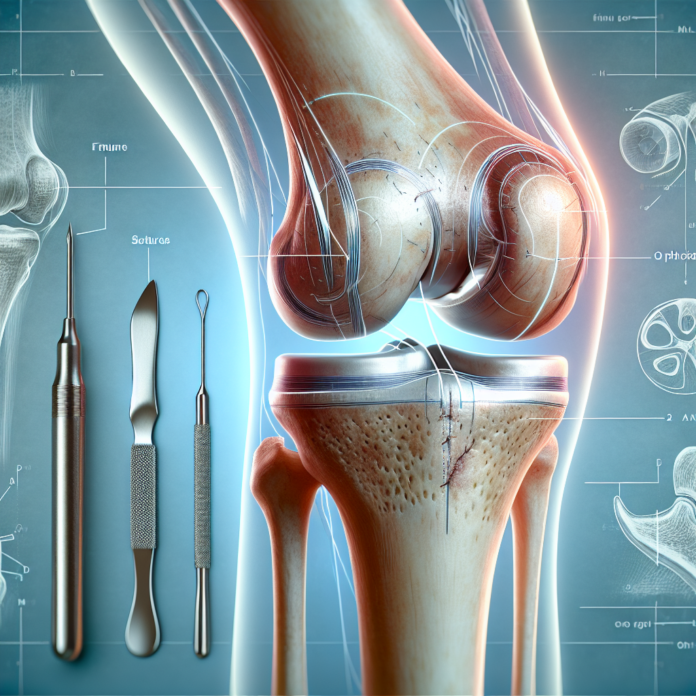
When discussing fractures of the medial femoral condyle, it’s important to first understand the anatomical context. The femoral condyles are the rounded ends of the femur or thigh bone that articulate with the tibia at the knee joint. Imagine them as the knuckles at the end of the thigh bone, connecting seamlessly with the tibia to allow for smooth and flexible movement. The medial femoral condyle, in particular, is located towards the midline of the body and plays a crucial role in the knee’s range of motion and weight-bearing function.
Occurrence and Causes
Interestingly, fractures of the medial femoral condyle are more prevalent in children than in adults. In children, bones tend to be less robust than the associated tendons and ligaments, leading to a higher likelihood of fractures under mechanical stress. In adults, ligaments are typically weaker than the bones, which can make ligament injuries more common. However, when a medial femoral condyle fracture does occur in an adult, it’s usually the result of a high-impact trauma, such as a car accident or a fall from a significant height.
Potential Complications
The nature of these fractures often involves a shearing pattern, which can cause a misalignment of the bone and the overlying cartilage. This misalignment poses a serious threat to the knee’s functionality and integrity. If left untreated, it can result in a “step-off” in the articular cartilage—a disruption that could lead to uneven wear and tear. Over time, this can cause degenerative changes, including the development of osteoarthritis, due to the abrasive nature of the misaligned surfaces.
Surgical Intervention
Due to these potential complications, surgical intervention is often recommended to realign the bone and restore the natural anatomy of the knee joint. The surgical procedure typically involves placing a plate and screws to securely hold the bone fragments in their correct position. This realignment is crucial not only for healing but also for maintaining long-term joint health.
Recovery Process
Once surgery is performed, the path to recovery is careful and measured. Healing time can vary significantly depending on factors such as the patient’s age, general health, and the precise nature of the fracture. Generally, a period of three to four months is required for the bone to heal adequately. During this time, the patient must adhere to specific weight-bearing restrictions to prevent any displacement of the fracture that could hinder recovery or necessitate further surgery.
A key component of the recovery process is the gradual reintroduction of weight-bearing activities. The orthopedic surgeon will typically set a timeline that allows for partial weight-bearing as the bone becomes more stable, usually beginning around six to eight weeks post-surgery. For some patients, weight-bearing restrictions might last up to twelve weeks, and it’s crucial that the patient follows these guidelines closely to promote optimal healing.
Another important consideration in the recovery is the impact on daily activities, including work. The timing of a return to work largely depends on the nature of the patient’s occupation. Those with sedentary jobs may resume work sooner, sometimes within a few weeks of surgery, provided the work environment accommodates the necessary mobility aids like crutches or a walker. Conversely, those whose jobs entail standing, walking, or physical activity may require a longer period of recovery and rehabilitation before they can return to their regular duties.
Rehabilitation
Rehabilitation post-surgery is tailored to each patient’s needs and will often involve physiotherapy to restore strength and flexibility to the knee. Exercises are designed to be progressively challenging and aim to improve the range of motion and muscle strength around the knee joint.
Exploring Alternatives
While surgery is often necessary for ensuring the proper healing of a medial femoral condyle fracture, it is not the only option available. For individuals interested in exploring non-surgical treatments or complementary therapies, consulting with a healthcare professional who specializes in regenerative medicine may provide alternatives such as physical therapy, rehabilitation exercises, or even newer techniques like platelet-rich plasma (PRP) treatments.
Ultimately, understanding the nature of a medial femoral condyle fracture, the surgical procedures involved, and the nuances of a successful recovery process is essential for both patients and practitioners. The recovery process is not merely about healing the bone but ensuring that the knee joint returns to its full functional capability, thereby enhancing the patient’s overall quality of life.
Dr. David Guyer discusses medial femoral condyle fractures, surgery need, and recovery. Learn about this rare injury, often in children, and healing processes.
