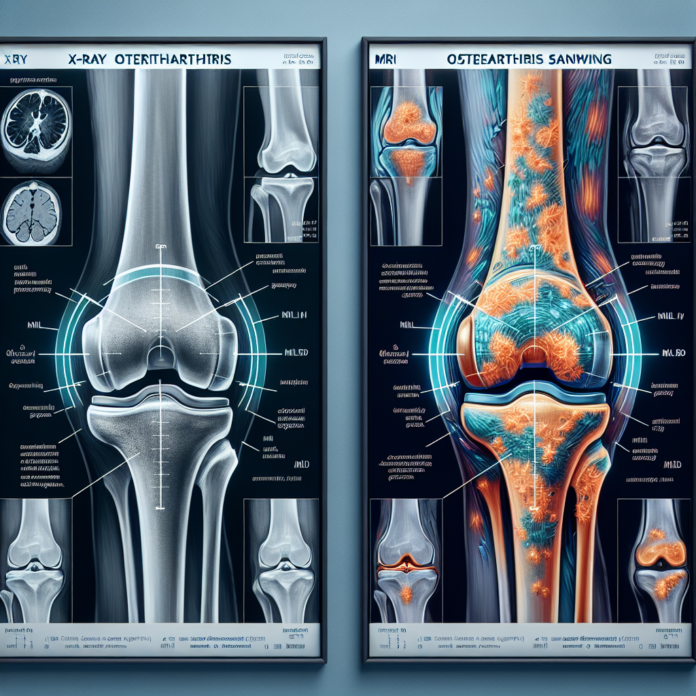
Osteoarthritis is a common ailment that affects millions of people worldwide, causing joint pain and reducing quality of life. When it comes to diagnosing and assessing the severity of this condition, two of the most commonly used imaging tests are X-rays and MRIs. But how do you know which one is right for you? In this blog, we will delve into the pros and cons of each test, shedding light on how they each capture different aspects of osteoarthritis. Based on insights from Dr. David Guyer, a triple board-certified orthopedic surgeon and expert in sports, anti-aging, and regenerative medicine, we’ll explore why one doctor may recommend an X-ray while another advises an MRI, and how other factors play into the decision of whether a joint replacement is necessary. Join us as we unpack the intricacies of these diagnostic tools and help you make informed choices about your osteoarthritis treatment plan.
X-rays: A Staple in Diagnosing Osteoarthritis
When dealing with osteoarthritis, one of the first steps in managing the condition is understanding its severity and how it affects your joints. X-rays and MRIs are two diagnostic tools frequently used in this process, each with its unique strengths and limitations. Understanding these differences can help you make a more informed decision in collaboration with your healthcare provider.
X-rays have long been a staple in diagnosing bone-related conditions, including osteoarthritis. One of their primary advantages is their ability to show the bones and the spaces between them clearly. This is crucial because osteoarthritis is often characterized by the narrowing of the joint space, which indicates the wearing down of cartilage. X-rays are frequently performed in orthopedic settings, where they may include weight-bearing images.
Pros of X-rays:
- Clear Bone Visualization: X-rays are excellent at visualizing bone and can clearly show bone spurs and the narrowing of joint spaces.
- Weight-Bearing Images: This capability allows doctors to see how your bones behave under the pressure of your body weight, which can be more telling than an image taken without load.
- Cost-Effective: Typically, X-rays are less expensive than MRIs, which can be a consideration for those without extensive health insurance coverage.
MRIs: Seeing Beyond the Bones
Despite their usefulness in visualizing bone structure and joint space, X-rays have limitations. They cannot visualize soft tissues like cartilage, ligaments, or tendons, which are also affected by osteoarthritis. This is where MRIs come into play.
MRIs provide a comprehensive view of not just the bones, but also the soft tissues surrounding them. This can give a more complete picture of joint health.
Pros of MRIs:
- Detailed Soft Tissue Imaging: MRIs can show the condition of cartilage, identifying thinning, divots, or holes, alongside healthy versus degenerated tissue for comparison.
- Comprehensive Joint Assessment: They can also visualize tendons, ligaments, and the meniscus, providing a fuller picture of joint health.
Cons of MRIs:
- Lack of Weight-Bearing Perspective: Without the load-bearing factor, MRIs sometimes underestimate the extent of arthritis.
- Higher Cost: The expense can be a deterrent for some patients.
Making the Right Choice for Your Health
Both imaging techniques have their places in diagnosing osteoarthritis, with each providing valuable insights. However, neither can single-handedly dictate the need for treatments such as joint replacement surgery. The decision to proceed with a knee or hip replacement involves considering various factors beyond just imaging results. These factors often include:
- Severity of Pain: How significantly does the pain affect daily activities and quality of life?
- Previous Treatments: Have non-invasive therapies like physical therapy or medications been tried?
- Functional Limitations: What activities can the patient no longer perform due to joint degeneration?
These considerations emphasize that imaging results are only one part of a larger puzzle in managing osteoarthritis. The role of a healthcare provider is to integrate imaging findings with clinical evaluation and patient history to tailor a treatment plan that best suits the individual’s needs.
Choosing between an X-ray and an MRI should be an informed decision made with your healthcare provider, considering the specific aspects of your condition that need to be visualized and the potential cost implications.
For those seeking more information and alternative options to surgery, exploring resources or consulting with a specialist can provide additional insights. Dr. David Guyer has highlighted resources like his eBook “The Arthritis Solution” for patients wanting to delve deeper into understanding osteoarthritis and its management.
By equipping yourself with knowledge and understanding the role each diagnostic tool plays, you can better engage in discussions about your health and the most appropriate course of action for your condition. Whether you lean towards an X-ray, an MRI, or both, the key is that the choice should support a holistic view of your health needs and treatment goals.
