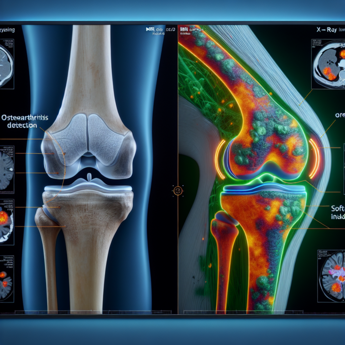
On the quest to accurately diagnose and evaluate the severity of osteoarthritis, patients often wonder whether X-rays or MRIs are the more effective tool. In this blog, we’ll dive into the strengths and limitations of each imaging method, particularly when it comes to illustrating bone damage, cartilage health, and the presence of bone spurs. Dr. David Guyer, a triple board-certified orthopedic surgeon, will guide us through the nuances of these diagnostic tools, highlighting how they contribute to assessing the need for interventions such as hip or knee replacements. Understanding the role of gravity, body weight, and imaging in the interpretation of osteoarthritis can empower patients to navigate their treatment options more effectively. Whether you’re considering these tests or exploring alternatives to surgery, this comprehensive analysis will equip you with critical insights into managing osteoarthritis.
X-Rays in Osteoarthritis Diagnosis
X-rays and MRIs are cornerstone imaging tools used to diagnose and assess the severity of osteoarthritis. Each method offers unique insights into the condition, yet neither stands as the sole determinant for interventions like joint replacement. Here, we break down the intricacies of how these diagnostic tools work and what they reveal about osteoarthritis.
X-rays are a primary diagnostic tool due to their ability to provide clear images of the bone structure. They measure the space between bones, which indicates cartilage health because X-rays visualize bones and not soft tissues.
- Advantages:
- X-rays are excellent for visualizing bone positioning and revealing bone spurs, which are direct indicators of joint use and damage.
- Weightbearing X-rays, taken while a patient stands or applies pressure on the joint, show the actual condition under gravitational stress. This is crucial because it simulates real-life conditions, making it easier to determine the true severity of bone-on-bone contact.
- Limitations:
- Since X-rays only capture the bone and not soft tissues, they cannot directly show cartilage erosion or tears.
MRI Advantages and Limitations
MRI scans offer a different perspective by imaging the soft tissues in and around the joints. They provide a detailed view of cartilage, meniscus, ligaments, and other soft tissues that play a significant role in joint function.
- Advantages:
- An MRI can show details of the cartilage, highlighting thinning or holes, giving a clear picture of the extent of cartilage damage.
- This imaging technique can identify other structural damages, such as ligament tears or meniscal injuries, which might be contributing to joint pain and dysfunction.
- Limitations:
- Unlike X-rays, an MRI is typically conducted while the patient is lying down. This absence of gravity and body weight can sometimes lead to an underestimation of the arthritis severity.
- MRIs are more expensive and time-consuming compared to X-rays, which might not always be feasible for frequent monitoring.
Complementary Role of X-rays and MRIs
Both X-rays and MRIs provide valuable information but from different perspectives. They are complementary tools in the diagnostic toolkit rather than competitors.
- For a comprehensive assessment, understanding the interaction between the solid bone structures (via X-rays) and the soft tissue conditions (via MRI) provides a holistic view of joint health and deterioration.
- Practical Implications:
- The decision to proceed with a hip or knee replacement doesn’t solely hinge on imaging results. Doctors consider multiple factors, including the severity of the pain, impact on daily activities, and patient-specific health concerns.
- Imaging results are one part of a broader diagnostic process that includes clinical evaluation and patient-reported symptoms.
Navigating Treatment Decisions
Determining the appropriate intervention for osteoarthritis is a complex decision-making process. The imaging results are integrated with patient history, physical examination, and patient preferences.
- Patient-Centric Approach:
- Treatment plans should respect the patient’s lifestyle, activity level, and personal goals. For some, preserving the joint as long as possible is a priority, whereas others may prioritize pain relief even if that means undergoing a joint replacement.
- Alternatives to Surgery:
- There are various non-surgical options available for managing osteoarthritis, including physical therapy, pharmacotherapy, and newer regenerative medicine techniques.
- Understanding all available treatment options, including lifestyle modifications and emerging therapies, is crucial for patients looking to delay or avoid surgery.
Conclusion
In conclusion, both X-ray and MRI have distinct but complementary roles in diagnosing and assessing osteoarthritis. While X-rays provide essential insights into bone structure and alignment, MRIs offer a detailed look into the soft tissues that X-rays cannot show.
For effective management of osteoarthritis, it is crucial to integrate the insights from both imaging modalities with clinical symptoms and patient preferences. Empowering patients to understand these diagnostic tools helps them make informed decisions about their treatment journey, whether that involves surgery or exploring other therapeutic avenues.
