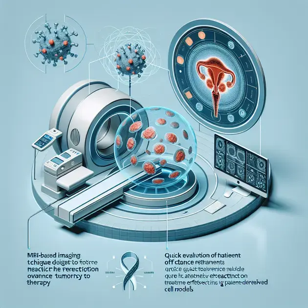
An MRI-based Imaging Technique Predicts Ovarian Cancer Treatment Response and Efficacy
Ovarian cancer is a devastating disease that affects thousands of women worldwide. While various treatment options exist, determining the most effective treatment strategy for each patient remains a challenge. However, a recent study has shown promising results in using MRI-based imaging techniques to predict the response of ovarian cancer tumors to treatment and to rapidly evaluate treatment efficacy in patient-derived cell models.
The study, conducted by a team of researchers, utilized advanced MRI imaging to assess ovarian cancer cell samples obtained from patients. By closely monitoring the changes in the tumors’ microenvironment and the response of tumor cells to treatment, they were able to accurately predict the outcome of various treatment options.
The MRI-based imaging technique relies on analyzing distinct features of the tumor microenvironment, such as blood flow, oxygen levels, and cellularity. By monitoring these parameters before and after treatment, researchers observed significant changes in the tumors’ characteristics, allowing them to gauge the treatment response.
The team successfully identified specific imaging signatures associated with treatment resistance and sensitivity, enabling them to differentiate between tumors likely to respond well to therapy and those that may not. This valuable information could potentially guide clinicians in selecting appropriate treatment options, avoiding unnecessary delays in identifying the most effective approach.
Moreover, the MRI-based imaging technique proved to be a rapid and non-invasive method for assessing treatment efficacy. Traditional response evaluation methods often rely on repeated biopsies or invasive procedures, which are not only time-consuming but can also cause discomfort to patients. In contrast, MRI imaging allows for real-time monitoring of tumor response, providing clinicians with immediate feedback on treatment effectiveness.
While this study provides encouraging evidence of the potential clinical applications of MRI-based imaging in ovarian cancer treatment, further research is needed to validate these findings in larger patient populations. Nevertheless, the prospect of utilizing this imaging technique to predict treatment response and assess therapy efficacy holds great promise for improving patient outcomes in ovarian cancer.
In conclusion, the groundbreaking research utilizing an MRI-based imaging technique offers a novel approach to predicting the response of ovarian cancer tumors to treatment and evaluating treatment efficacy. This non-invasive method has the potential to revolutionize ovarian cancer treatment by enabling personalized therapeutic strategies and real-time monitoring of treatment response. Further studies and clinical trials are necessary to fully establish the clinical utility of this technique, but its potential impact cannot be underestimated in the fight against ovarian cancer.